In Vivo Staining and Identification of Plant Cells
1. Principle
Living dyeing is a technique for dyeing living cells with a dye solution that is not harmful to plants. Neutral red is one of the commonly used living dyes. It is a weakly alkaline pH indicator with a color range between pH 6.4-8.0 (from red to yellow).
In a neutral or slightly alkaline environment, the living cells of the plant can absorb a large amount of neutral red and excrete into the vacuole, because the vacuole generally exhibits an acidic reaction. Therefore, the neutral red that enters the vacuole dissociates and releases a large amount of cations to appear cherry red. In this case, the protoplast and cell wall are generally not colored; dead cells are not maintained in the vacuole due to denaturation and solidification of the protoplast. Therefore, after dyeing with neutral red, no bubble coloring occurs. On the contrary, the neutral red cations combine with the negatively charged protoplast and nucleus to stain the protoplast and nucleus.
2. Instruments and appliances
Microscope; small petri dish; glass slide; cover glass; single-sided blade; pointed tweezers; alcohol lamp; match; wipe lens paper;
3. Reagents
0.03% neutral red solution; 1mol / L potassium nitrate solution.
Four, methods
1. Choose onion bulbs (or the base of green onion pseudostalks) and wheat leaves as materials.
2. Cut off a younger onion scale, cut into small pieces of about 0.5cm2 on the inside of the scale with a single-sided blade, and gently tear off the small pieces of the inner epidermis with pointed tweezers to put into the neutral red solution Stain (note that the inside of the epidermis should be down). If you use wheat leaves as the material, you can lay the back of the leaves flat on a glass slide, put the glass slide in a Petri dish containing a small amount of water, press the leaf flat with the left hand, and use the blade from one direction Gently scrape off the epidermis and mesophyll parts, leaving only the transparent upper epidermal cells. Be careful when scraping only a small amount of mesophyll cells. Too much force will easily damage the epidermal cells, and even only a layer of cell wall will be left. Too light will leave too many mesophyll cells, affecting the observation. In addition to wheat, other gramineous plants can also use this method to prepare epidermal cells, and cut the scraped material into small pieces of about 0.5 cm2.
3. Put the prepared cuticles of onion bulbs or the epidermis of wheat leaves into 0.03% neutral red solution for dyeing for 5 ~ 10min, take out 1 ~ 2 pieces, rinse a little in distilled water, and drop a drop on the slide Distilled water, carefully flatten the slide onto a glass slide, cover the glass, and observe under a microscope, you can see that the cell wall is stained red, but the protoplasts and vacuoles are not stained, because the distilled water is acidic and weakly acidic Under negative conditions, the cell wall is negatively charged and the adsorption of dye cations results.
4. Take a few of the biopsy preparations from step 3 and put them in tap water with a pH slightly higher than 7.0 for 10-15 minutes, and then put them on a glass slide for microscopy. The cell walls will be discolored, but the vacuoles will be stained deep Red, this is because when the solution pH is higher than 7.0, the dissociation of neutral red molecules is very weak, mainly in molecular state, not easy to be adsorbed by the cell wall, but easier to enter the vacuole through the plasma membrane and vacuole membrane, The plant sap is mostly acidic, and the neutral red that enters the vacuole dissociates, dyeing the vacuole cherry red. At this time, the nucleus and protoplasts are not stained.
In order to confirm the neutral red staining site, the above-mentioned onion inner skin staining sheet may be immersed in a 1 mol / L potassium nitrate solution for about 10 minutes, and then taken out for observation. Because potassium nitrate can cause the protoplasts to swell strongly, "capped wall separation" occurs, so that the colorless and transparent protoplasts can be clearly distinguished from the red-stained vacuoles.
5. Place the live slice in step 4 on the flame of an alcohol lamp and heat it slightly to kill the cells. Observe under a microscope to see that the protoplasts coagulated into an uneven gel and stained red with the nucleus .
6. Look carefully in the biopsy preparation, you may see some dead cells, and their nucleus is red because of neutral red staining, clearly distinguishable.
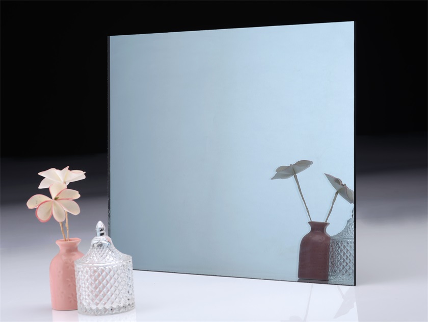
A Silver Mirror is a type of glass mirror. Silver mirrors are commonly known as waterproof mirrors, mercury mirrors, silver-plated mirrors on glass surfaces, glass mirrors, mirror glass, etc. Silver mirrors are widely used in furniture, handicrafts, decoration, bathroom mirrors, cosmetic mirrors, optical mirrors and car rearview mirrors. When storing mirrors, they should not be stacked with alkaline and acidic substances, and should not be stored in a humid environment.
In addition, we also sell Silver Mirror glass, silver mirror commonly known as waterproof mirror, mercury mirror, silver-plated mirror on glass surface, glass mirror, mirror glass, etc. Silver mirrors are widely used in furniture, handicrafts, decoration, bathroom mirrors, cosmetic mirrors, optical mirrors, and car rearview mirrors.

Clear Silver Mirror,Clear Silver Mirror Bar,Clear Silver Mirror Bathroom,Clear Silver Mirror Lens
Dongguan Huahui Glass Manufacturing Co.,Ltd , https://www.antiquemirrorsupplier.com