1. Organizational processing
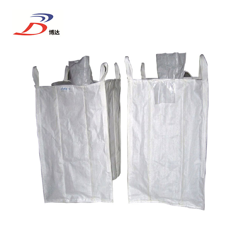
This category of pp jumbo bags are the most common Big Bag available. Jumbo Plastic Bags contains a load capacity that ranges between 500kgs to 2000kgs. Bulk Bag Volume dimensions alter depending upon the preferences and needs of the customer. Pp Container Bag is used to transport non-flammable dry substances that are in powdery form
No.
Item
Specification
1
Size
85cm*85cm*90cm/90cm*90cm*100cm or customized
2
Body construction
4-panel/U panel/Circular panel/Tubular panel/rectangular type
3
Top
Open mouth/skirt mouth/ filling spout
4
Bottom
Flat /discharge spout
5
Loop type
side seamed /cross corner/double stevedore with 2-4 belts
6
Printing type
one or two side with 1-3 color off set color
7
Optional parts
document pouch/label/rings/PE liner
8
SWL
5:1/3:1/6:1
9
Loading capacity
500kg to 3000kg
10
Color
white, yellow, blue or customized
11
Fabric weight
100g/m2 to 240g/m2
Jumbo Plastic Bags,U-Pannel Jumbo Bag,Jumbo Duffle Bag,Pp Container Bag Shijiazhuang Boda Plastic Chemical Co., Ltd. , https://www.ppwovenbag-factory.com
Proper tissue processing is a prerequisite for good immunohistochemical staining, and an internal factor that determines the success or failure of staining. In the process of preparing tissue cells, not only the integrity of tissue cells but also the antigenicity of tissue cells must be maintained. Damage or diffuse, prevent tissue autolysis. If there is autolysis and necrotic tissue, the antigen has been lost. Even with very sensitive detection antibodies and superb technology, it is difficult to detect the desired antigen. Instead, it is often due to tissue necrosis or squeeze during the preparation of the knife. False positive results are prone to appear in the above areas.
1) Organize materials and fix in time
The timely collection and fixation of tissue specimens is the key first step in immunohistochemical staining, which is to effectively prevent tissue autolysis and necrosis, and the start of antigen loss. The isolated tissue should be collected as soon as possible, preferably within 2h, when used for sampling The knife should be sharp. Cut the tissue with a single knife. Do not cut and pull the tissue repeatedly, causing tissue compression. The size of the tissue block should be moderate, generally 2.5cm × 2.5cm × 0.2cm. Remember to use a large tissue block when collecting materials. The principle of not being thick, (that is, the area of ​​the tissue block can be as large as 3cm × 5cm, but the thickness of the tissue block must not exceed 0.2cm, otherwise it will not be conducive to the uniform fixation of the tissue). The fixative fluid quickly penetrates into the tissue so that the tissue protein can quickly solidify within a certain time. Thus, the antigen and tissue cell morphology are preserved intact.
For the selection of fixative fluid, in principle, the corresponding fixative fluid should be selected according to the tolerance of the antigen, but unless it is a special scientific research project, it is difficult to do this in routine pathological work, because both pathological diagnosis and differential diagnosis It is based on routine HE pathological diagnosis to decide whether to perform immunohistochemical staining, and the conventional tissue treatment of HE staining is performed with 10% neutral buffered formalin or 4% buffered paraformaldehyde 4 times the tissue volume Tissue fixation, with its strong permeability, fixes the effect of the tissue evenly, but the tissue fixation time is preferably within l2h, and the general fixation time should not exceed 24 hours. With the extension of a fixed time, the detection intensity of tissue antigens will gradually decrease.
2) Tissue dehydration, transparent, wax dipping
After the tissue is fixed, it is dehydrated, transparent, dipped in wax and embedded. The principle of grasping is that the dehydration and transparency should be sufficient but not over, the wax dipping time should be sufficient, and the temperature should not be high, otherwise it will make the tissue hard and brittle and make tissue slicing difficult. Even if it can be sliced, because the tissue is hard and brittle, the slice cannot be intact and smooth. It is very easy to take off the film during the staining process, which is unfavorable for the location and background of the immunohistochemical staining antigen. Therefore, the time for anhydrous alcohol dehydration and xylene transparency should not be too long. Normal size tissues are dehydrated with anhydrous alcohol for lh × 3 times Toluene can be transparent lh × 2 times, and the temperature of the dipping wax and embedded paraffin should not exceed 65 ℃.
2. Slice
After the tissue is well processed, the glass slide should be processed before sectioning. Due to the variety of antigens we detect, due to the complicated staining procedure and long time, some antigens need to be repaired by various antigens, such as microwave , High pressure, water-soluble enzymes, etc., if the slides are not handled well, it will be easy to cause off-chip. In order to ensure the normal progress of the immunohistochemistry experiment, it is required to properly handle the slides before the placement and must be cleaned. Clean the slides with adhesive to prevent them from falling off.
1) Poly-L-Lysine (Poly-L-Lysine)
Generally, 0.5% polylysine with a molecular weight of about 30,000 is the best. It can also be diluted with 1:10 deionized water in its concentrated solution sold by the reagent company. The method is to soak the slides, pour out the remaining liquid, and bake them in a 60 ° C incubator for use. The advantage of this method is that it can be used in a variety of histochemical, immunohistochemical and molecular detection applications, and the paste effect is the most Good, but the price is slightly more expensive.
2) Gelatin chromium potassium sulfate method
Dissolve 2.5g of gelatin in 500ml of distilled water by heating, dissolve and cool it down, add 0.25g of chromium potassium sulfate and mix well to dissolve it. The method is to soak the slides for 2 minutes, take out the controlled liquid into the incubator and dry it for use. This method is cheap and simple, and can be used in any experiment, especially for large-scale use, but it should be noted that if the liquid becomes blue or viscous, it is disabled.
3) APES (3-aminopropyl-ethoxysilane)
This method must be used now. Put the washed slide into APES diluted with 1:50 acetone, soak for 20s, remove it and stop it, then brush it with acetone or distilled water to dry the unbound APES and dry. The slides bonded in this way should be baked vertically and not copied flatly, otherwise bubbles will easily appear in the tissue slices.
The slices must be kept sharp, the slices should be thin and flat, without wrinkles, and without marks. If the above problems occur, the slices will show false positives during immunohistochemical staining. The slice thickness is generally 3 ~ 4μm. The slices are kept in a 60 ° C incubator overnight. Note that the temperature of the roasted slices should not be too high, otherwise the tissue structure will be easily destroyed, and the phenomenon of antigen localization will be diffused.
3. Immunohistochemical staining
The S-P immunohistochemical staining kit uses a mixture of biotin-labeled secondary antibody and peroxidase linked to streptavidin and substrate pigment to determine antigens in cells and tissues.
S-P immunohistochemical staining steps:
1) After dewaxing and hydrating paraffin sections, rinse with PBS (pH 7.4) three times for 3 minutes (3 × 3 ′) each time.
2) According to the requirements of each antibody, repair the tissue antigen accordingly.
3) Add 1 drop or 50ul peroxidase blocking solution (reagent A) to each slice to block the activity of endogenous peroxidase and incubate at room temperature for 10 minutes.
4) Rinse 3 × 3 'with PBS.
5) Shake off the PBS solution, add 1 drop or 50ul of non-immune animal serum (reagent B) to each slice, and incubate at room temperature for 10 minutes.
6) Remove the serum, add 1 drop or 50ul of primary antibody (user-selected) to each slice, and incubate at room temperature for 60 minutes or overnight at 4 ° C. It is recommended to refer to the instructions for each antibody.
7) Rinse 3 × 5 'with PBS.
8) Shake off the PBS solution, add 1 drop or 50ul of biotin-labeled secondary antibody (reagent C) to each slice, and incubate at room temperature for 10 minutes.
9) Wash 3 × 3 'with PBS.
10) Remove the PBS solution, add 1 drop or 50ul of Streptomyces avidin-peroxidase solution (reagent D) to each slice, and incubate at room temperature for 10 minutes.
11) Rinse 3 × 3 ′ in PBS.
12) Remove the PBS solution, add 2 drops or 100ul of freshly prepared DAB or AEC solution to each slice, observe under the microscope for 3-10 minutes, the positive color is brown or red.
13) Rinse with tap water, counterstain with hematoxylin, differentiate with 0.1% HCL, rinse with 0.1% ammonia or PBS to return to blue.
14) If the color is developed with DAB, the slices are dehydrated and dried with gradient alcohol (xylene transparent), and neutral gum is mounted; if the color is developed with AEC, the slices cannot be dehydrated with alcohol, and the tablets are directly sealed with water-based sealing
